Photoreceptor Signaling
We study photoreceptors in both health and disease, including electrical and biochemical signaling in rod and cone outer segments, the delicate interdependence of the photoreceptors with the Müller cells and RPE for healthy physiology, and rod circuitry in the retina.
I. Phototransduction
Rods are so sensitive that they can signal the absorption of a single photon, and we can use these electrical responses to understand how a single activated rhodopsin generates an amplified biochemical response.
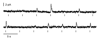
A rod’s quantized responses to dim flashes – sometimes one photon gets absorbed, sometimes two, sometimes zero!
Recordings by Dr. Owen Gross. All rights reserved. Burns Lab, UC Davis.
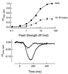
From the electrophysiological responses to both dim and bright flashes, we can determine the active lifetime of photoexcited rhodopsin, the lifetime of the transducin-PDE complex, the basal rate of cGMP turnover, and calcium exchange currents in a wide variety of mouse models. We also use computational modeling to test our understanding and mechanistic interpretations.
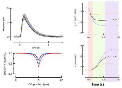
From Gross, Pugh, and Burns Biophys J (2012) and Gross, Pugh, and Burns Neuron (2012). All rights reserved. Burns Lab, UC Davis.
II. Rod and Cone Physiology and Homeostasis
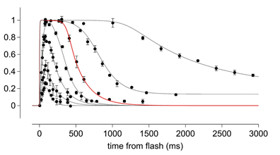
Normal photoreceptor signaling requires continual support of the RPE and Müller glia. For these reasons we are currently using both electrical and optical methodologies for studying both rods and cones in vivo.
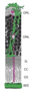
A. Light micrograph of a section of outer retina with a schematized Müller cell (green) and sheet of RPE intimately surrounding a rod (purple). Courtesy Dr. Gabriel Peinado. All rights reserved. Burns Lab, UC Davis.
B. Family of flash responses of rods obtained in vivo using the paired flash EGR paradigm. Adapted from Peinado et al., (2017) J Gen Physiol.
We are also developing physiological metrics to assess photoreceptor stress, which in turn guides our experiments investigating why exaggerated phototransduction signaling causes cell death.
III. Retinal Circuitry
Rods and cones piggyback on many of the same types of bipolar cells, which makes teasing apart the relative contributions of rods and cones to signaling within any given downstream cell type challenging.

A. The primary rod pathway (green), through which the single photon responses are collected, is also quite capable of light adaptation. Diagram courtesy Dr. Chris Fortenbach. All rights reserved. Burns Lab, UC Davis.
B. Figure of wild-type rod bipolar recordings in dark-adapted or light-adapted retinas from Long et al., 2013 Plos One.